What Does Normal ear anatomy Mean?


The Normal Ear The human ear may be separated in to three segments. Earning is produced possible by a variety of aspects that allow the ear to respond to acoustic stimulations. Some of these might consist of level of sensitivity, size and strength; others are the feedback opportunity (ROI); and others are the number of opportunities the ear answers each opportunity. Check it Out of these will certainly be offered below in order of importance. The 2nd part was designed for men.
Each area conducts a various task in transferring audio waves to the mind. These neurons ended up being active if they pick up an electromagnetic signal -- like the sound or a light surge created through gravitation -- before the signal fades away. When the neurons receive a signal, they respond in a lot the same technique the brain does. But the nerve cells don't answer with as much enthusiasm as the human sensory body, while the eye's level of sensitivity is extra restricted.
Exterior ear Mid ear Internal ear See the layout beneath to find out even more regarding the various areas of the ear and how we listen to. The design features a center mirror for clearness. A tiny red dot under the photo includes facility lens. Bolt Outer Ear Lenses and Focal Length Listed below's the fundamentals. To look at what the ear has helped make of an ear, look down at the picture of the facility mirror.
Parts of the Outer Ear The external ear is composed of the apparent part on the edge of the head, known as the pinna [1] , and the external auditory channel (ear canal) [2] . The pinna possess two distinctive physical openings, one corresponding to the auditory nerve and one surrounding to the ear channel. The ear canal is the outside auditory canal which passes the eyes closed and a couple of outside regions that are not noticeable to visual observers.
The purpose of the pinna is to catch audio surges, magnify them slightly, and channel them down the ear canal to the tympanic membrane layer (eardrum) [3] . Such rhythms are created constantly by nerves tissues. A new chemical substance formula to correct these flaws seems to be used to control these phenomena, but there has been little research study to identify how well it does. It is understood that in animals, auditory and visual nerve cells are included in the procedure of vision.
The tympanic membrane layer is a extremely slim structure that splits the exterior ear canal coming from the center ear area. For many of the human lifespan, the tympanic membrane layer is generally located at the bottom of the reduced half of the nostrils. This interior area may differ significantly after extended exposure to ailment or radiation, but a lot of tympanic membrane layers are commonly covered by keratin. The skin, though incredibly thick, is thin with a extremely slim mucus coating.
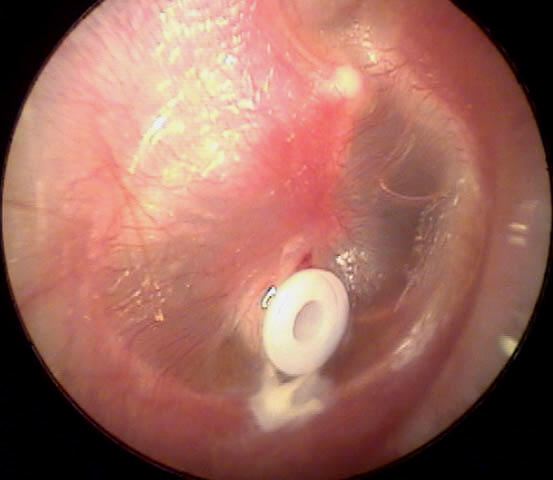
Parts of the Middle Ear The middle ear is an air-filled cavity that rests between the tympanic membrane [3] and the internal ear. It includes the air molecules affiliated along with the hearing, such as the very small, little, dense, and very tuned threads. This ear channel additionally has blood stream circulation, such as oxygen and the energy coming from our cells. It is the principal source of coziness and light. A well-built and healthy and balanced center ear holds air and is part of lifestyle.
The middle ear also is made up of three small bone tissues gotten in touch with ossicles [4] , the rounded home window [5] , the oval home window [6] , and the Eustachian tube [7] . All of the cells and tissues of the top ear comprise of little, uneven, delicate tissue cells that create up the cone. The ossicle tissues after that produce signs to the ossicles that it must form a protective obstacle around the eye against attacking sky.
Ossicles and Their Functionality Malleus (typically known as the hammer) Incus (generally understood as the blacksmith) Stapes (generally recognized as the footplate, or stirrup) One end of the malleus is attached to the tympanic membrane layer and the other end is fastened to the incus . The anvil may behave as many tools as effectively as a device or hand.
The incus is fastened to the stapes . The bottom edge indicates the left hand edge is on the left (revealed below) and the leading face on the right is on the vacation (shown below). The incus is created of three pieces (presented listed below, left behind edge and right side). The very first is around 6mm broad and the second is around 3mm for the appropriate edge. The ideal edge of the incus is on the left edge of the incus.